Phantom & Duplex IQI
Test Phantom for Qualification of Image Plate Scanner Systems (CR)
CR Phantom can test all relevant parameters of CR Scanner systems including basic spatial resolution, unsharpness, contrast, MTF, laser beam jitter, scanner slipping and shading. These tests demanded and described in detail in standards ASTM E 2445-14, ISO 16371-1 and EN 14784-1 have to be performed periodically.
The CR Phantom exceeds these standards by including two Duplex wire type IQIs. Measuring points for shading correction are arranged in both axis directions – panorama and landscape. All required information is mapped on the image plate with a single X-ray exposure – the CR Phantom need not be rotated to generate the information of the second axis. This results in more accurate test scores and significant time savings.
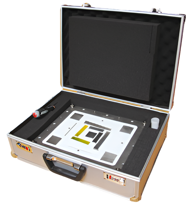
Specifications
-
T-Target – brass Laser beam jitter, MTF check,
-
Duplex wire type IQI
-
BAM snail
-
Converging line pair IQI
-
EL, EC, ER Measuring points
-
Cassette positioning locator
-
Homogeneous AL strip
-
Lucite plate
-
Cm/inch Ruler
-
Contrast sensitivity gauge
-
Dimensions: 350 x 430 x 19 mm
-
Laser beam jitter, MTF check, Blooming (Flare)
-
Basic spatial resolution, unsharpness
-
Central beam alignment
-
Line pair resolution
-
Shading correction
-
Position of cassette (image plate)
-
Scanner slipping, shading
-
Carrier plate
-
Linearity check
-
Contrast sensitivity check
-
14“ x 17“ x 0.75“
Duplex Wire Type IQI according to ISO 19232-5 Total Image Unsharpness Gage according to ASTM E 2002
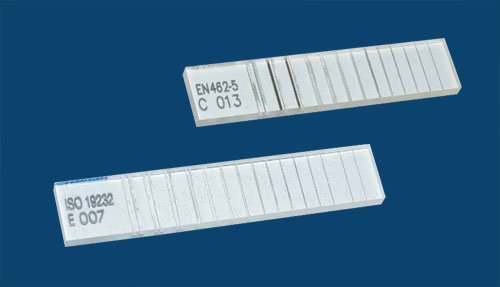
The Duplex IQI is used in many X-ray applications, especially in digital X-ray. Designed for evaluating total image unsharpness of film and additionally basic spatial resolution of digital images according to EN 13068 (Radioscopy), EN 14784 and ISO 13671 (CR – Computed Radiography with imaging plates), ISO 17636-2 (digital radiology of welds – flat panel detectors) or ASTM E 2597 (characterization of digital detector arrays). A new ISO standard is in preparation, describing possibility of using the Duplex IQI for determination of focal spot size of X-ray tubes.
In radiography the wire pair with largest diameter d has to be identified, which cannot be separated visually. The radiographic film may be magnified up to 4X.
In digital radiology the separation between wires (Dip) is evaluated. The wire pair with largest d, showing Dip less than 20 percent of the wire pair contrast, identifies total image unsharpness (U = 2d) and basic spatial resolution (SRb = d) of digital image.